Fascial Manipulation Boston
 Fascial Manipulation© is a manual therapy that has been developed by Luigi Stecco, an Italian physiotherapist from the north of Italy. This method has evolved over the last 35 years through study and practice in the treatment of a vast caseload of musculoskeletal problems.
It focuses on the fascia, in particular the deep muscular fascia, including the epimysium and the retinacula and considers that the myofascial system is a three-dimensional continuum. In recent years, via collaboration with the Anatomy Faculties of the René Descartes University, Paris, France and the University of Padova in Italy, Dr. Carla Stecco and Dr. Antonio Stecco have carried out extensive research into the anatomy and histology of the fascia via dissection of unembalmed cadavers. These dissections have enhanced the pre-existing biomechanical model already elaborated by Luigi Stecco (1,2) by providing new histological and anatomical data.
This technique presents a complete biomechanical model that assists in deciphering the role of fascia in musculoskeletal disorders.
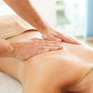
Precise Manipulation to Restore Movement
The mainstay of this manual technique lies in the identification of a specific, localised area of the fascia in connection with a specific limited movement. Once a limited or painful movement is identified, then a specific point on the fascia is implicated and, through the appropriate manipulation of this precise part of the fascia, movement can be restored.
While part of the fascia is anchored to bone, part is also always free to slide. The free part of the fascia allows the muscular traction, or the myofascial vectors, to converge at a specific point, named the vectorial Centre of Coordination or CC (5). The location of each CC has been calculated by taking into consideration the sum of the vectorial forces involved in the execution of each movement.
The six movements made on the three spatial planes are rarely carried out separately but, more commonly, are combined together to form intermediate trajectories, similar to the PNF patterns. In order to synchronize these complex movements other specific points of the fascia (often over retinacula) have been identified and, subsequently, named Centres of Fusion or CF.
Key Role in Peripheral Motor Control and Proprioception
Fascia is formed by undulated collagen fibres and elastic fibres arranged in distinct layers, and within each layer the fibres are aligned in a different direction. Due to its undulated collagen fibres, fascia can be stretched and, thanks to its elastic fibres, it can then return to its original resting state. Given that fascia adapts to muscle stretch, it is unable to transmit force like a tendon or an aponeurosis. If these histological and functional distinctions are not taken into consideration, then one can confuse fascia with aponeuroses or, likewise, confuse the deep fascia with the subcutaneous connective tissue (superficial fascia). Subcutaneous connective tissue forms a very elastic, sliding membrane essential for thermal regulation, metabolic exchanges and the protection of vessels and nerves, whereas the deep fascia envelops the muscles, and surrounds the muscle’s aponeurosis up to where it inserts onto bone.
It is hypothesised, that the richly innervated fascia (7) could be maintained in a resting state of tension due to the different muscular fibres that insert onto it. Due to this optimal resting state, or basal tension, of the fascia, the free nerve endings and receptors within the fascial tissue are primed to perceive any variation in tension and, therefore, any movement of the body, whenever it occurs.
Deep fascia is effectively an ideal structure for perceiving and, consequently, assisting in organizing movements. In fact, one vector, or afferent impulse, has no more significance to the Central Nervous System than any other vector unless these vectors are mapped out and given a spatial significance. In human beings, the complexity of physical activity is, in part, determined by the crossover synchrony between the limbs and a refined variability in gestures. Whenever a body part moves in any given direction in space there is a myofascial, tensional re-arrangement within the corresponding fascia. Afferents embedded within the fascia are stimulated, producing accurate directional information. Any impediment in the gliding of the fascia could alter afferent input resulting in incoherent movement.
It is hypothesised that fascia is involved in proprioception and peripheral motor control in strict collaboration with the CNS.
Therapeutic Implications
The fascia is very extensive and so it would be difficult and inappropriate to work over the entire area. The localisation of precise points or key areas can render manipulation more effective. An accurate analysis of the myofascial connections based on an understanding of fascial anatomy can provide indications as to where it is best to intervene. Any non-physiological alteration of deep fascia could cause tensional changes along a related sequence resulting in incorrect activation of nerve receptors, uncoordinated movements, and consequent nociceptive afferents. Deep massage on these specific points (CC and CF) aims at restoring tensional balance. Compensatory tension may extend along a myofascial sequence so myofascial continuity could be involved in the referral of pain along a limb or at a distance, even in the absence of specific nerve root disturbance. In clinical practice, cases of sciatic-like pain and cervicobrachialgia without detectable nerve root irritation are common (8).
Get More Information
Contact our Spine & Sports Injury Center team to learn more or book an appointment.
CONTACT US »

Fascial Manipulation Boston’s Back Bay, South End, Downtown, Financial District and Beacon Hill MA | (617) 247-2300
References
- Stecco L Fascial Manipulation for Musculoskeletal pain, Piccin, 2004
- Stecco L & C. Fascial Manipulation Practical Part, Piccin , 2009.
- Stecco C, Gagey O, Macchi V, Porzionato A, De Caro R, Aldegheri R, Delmas V.Tendinous muscular insertions onto the deep fascia of the upper limb. First part: anatomical study.Morphologie. 2007 Mar;91(292):29-37. PMID: 17574470 [PubMed – in process]
- Stecco C, Porzionato A, Macchi V, Stecco A, Vigato E, Parenti A, Delmas V, Aldegheri R, De Caro R.The Expansions of the Pectoral Girdle Muscles onto the Brachial Fascia: Morphological Aspects and Spatial Disposition.Cells Tissues Organs. 2008 Mar 19; [Epub ahead of print]PMID: 18349526 [PubMed – as supplied by publisher]
- Alessandro Pedrelli, Carla Stecco, Julie Ann DayTreating patellar tendinopathy with Fascial ManipulationJ Bodyw Mov Ther. 2009 Jan;13(1):73-80. Epub 2008 Jul 26.PMID: 19118795 [PubMed – indexed for MEDLINE]
- A. Stecco • V. Macchi • S. Masiero • A. Porzionato •C. Tiengo • C. Stecco • V. Delmas • R. De CaroPectoral and femoral fasciae: common aspects and regional specializationsSurg Radiol Anat 2008 DOI 10.1007/s00276-008-0395-5
- Stecco C, Gagey O, Belloni A, Pozzuoli A, Porzionato A, Macchi V, Aldegheri R, De Caro R, Delmas V.Anatomy of the deep fascia of the upper limb. Second part: study of innervation.Morphologie. 2007 Mar;91(292):38-43. PMID: 17574469 [PubMed – in process]
- Day JA, Stecco C, Stecco A.Application of Fascial Manipulation technique in chronic shoulder pain–anatomical basis and clinical implications.J Bodyw Mov Ther. 2009 Apr;13(2):128-35. Epub 2008 Jun 24. PMID: 19329049 [PubMed – in process]